


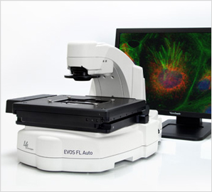
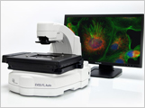
![]() > Life technologies > EVOS fl auto
> Life technologies > EVOS fl auto
 |
|
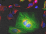
| Fluorescence, brightfield and phase contrast | |
| Patented LED light cube technology | |
| Supports up to 4 fluorescence channels simultaneously | |
| supports up to color and monochrome cameras | |
| ease-of-use with X/Y scanning stage, autofocus and | |
| flat-focus Z-stack | |
| Advanced imaging software (high-resolution mosaic tiling, | |
| multi-position well scanning, object counting, and time-lapse) | |
| 22" touch-screen LCD display, networking capability, | |
| and DVI output | |

Optics
- Infinity-corrected
- RMS-threaded objectives with 45 mm parfocal distance
Objective Selection
- 2X, 0.06 NA, 6.0 mm WD
- 4X Ph, 0.13 NA, 17.2 mm WD
- 4X Fl, 0.13 NA, LWD
- 10X Fl, 0.30 NA, 7.1 mm WD
- 20X Fl, 0.45 NA, 5.9 mm WD
- 40X Fl, 0.65 NA, 1.6 mm WD
- 60X Fl, 0.75 NA, 1.0 mm WD
- 4X Ph/Fl, 0.13 NA, LWD
- 10X Ph/Fl, 0.25 NA, LWD
- 20X Ph/Fl, 0.40 NA, LWD
- 40X Ph/Fl, 0.65 NA, LWD
- 60X APO Oil, 1.42 NA
- 50x Achro objective
- 100x Achro objective
- 100X Fl Oil, 1.28 NA (w/ Cleaning Kit)
- 20X Fl coverslip-corrected, 0.50 NA
- 40X Fl coverslip-corrected, 0.75 NA
LED Light Cube Selection
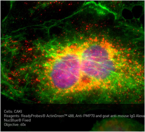
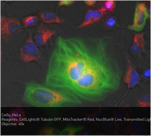
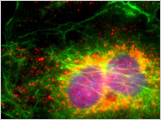
- DAPI (357 nm excitation, 447 nm emission)
- CFP (442 nm excitation,510 nm emission)
- GFP (470 nm excitation, 525 nm emission)
- YFP (500 nm excitation, 542 nm emission)
- RFP (531 nm excitation, 593 nm emission)
- Texas Red (585 nm excitation, 624 nm emission)
- CY5 (628 nm excitation, 692 nm emission)
- CY7 (731 nm excitation, 825 nm emission)
- QDots (all available wavelengths)
Illumination
- Patented LED Light Cubes (50,000+ hour life)
- Adjustable intensity
- Instant ON/OFF
- Easy install, no maintenance
Contrast Methods
- Fluorescence and transmitted light (brightfield or phase contrast)
Dimensions
- Height: 32.2 cm (12.7 in)
- Width: 34.3 cm (13.5 in)
- Depth: 47.2 cm (18.6 in)
Weight
- 20.0 kg (44.1 lbs)
Condenser
- 4-position turret for brightfield and phase contrast
- Slider with diffuser block and meniscus filters
Condenser Working Distance
- 60 mm
Stage
- automated X/Y scanning stage
- ravel range: 115mm x 78mm with sub-micron resolution
- Interchangeable vessel holders for most common shapes and sizes
- vessel holders lock down mechanism to fix sample in place during long scans
Focus mechanism
- automated focus mechanism with sub-micron resolution
LCD Display
- 22" high-resolution touch screen color monitor (also fully controllable via mouse), 1920 x 1080 pixel resolution
Cameras Available
- dual monochrome and color cameras
- monochrome camera : Sony® ICX445 monochrome CCD, 1/3" 1280 x 960 pixels, 1.3 megapixels
- Color camera: CMOS digital camera 1360 x 1024 pixels, 3.1 megapixels
Computer
- external PC
- 16 GB RAM running Windows® 7 Pro
- touch-screen monitor
Software
- Auto exposure : adjustment for optimal exposure times
- Autofocus : can be set up in several different modes to optimize speed and accuracy
- Automated optical system : automated switching of parfocal objectives, LED light cubes, and monochrome/color cameras
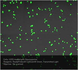
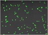
- Counting function : automated cell counting with watershed algorithm for easy GFP expression monitoring, live/dead assessment, and total cell count using NucBlue® Live
- Custom pseudocolor : choose display color for each cube (monochrome camera only)
- Freehand annotation tool : highlight areas/cells of interest using the touch-screen controls or mouse to draw freehand shapes
- Image editing : Counting, measurements, annotation tools, and image review; automatic overlay, select/deselect channels, brightness/contrast adjustments, and saving to USB or network
- Image stitching : capture multiple images with overlapping fields and use mosaic tiling to stitch a high-resolution image of a large area
- Image Review : bring in images for editing or object counting
- Manual exploration : define parameters optimal for your experiments
- Measure and annotate : add annotations and scale bar
- Quick Image Wizard : step-by-step guide to capture publication-quality multicolor images
- Reduced photobleaching : variable illumination control of digital LED light source and camera signal amplification helps dramatically reduce photobleaching
- ROI definition : define regions of interest (ROI) for scan routines
- Routines : create, save, and run repeat routines and experiments
- Scalebar : a scalebar can be added to a captured image via the System Basics tab
- Scan review : zoom in/out and pan, export the entire scan or only regions of interest
- Time lapse : use up to 8 beacons to seamlessly capture time-lapse movies on slides or microplates
- Z-Stack flat focus : collect a series of images; extract the most "in focus" pixels from each to create a single, focused image even from thick samples
Captured Images
- 16-bit monochrome TIFF or PNG (12-bit dynamic range)
- 24-bit color TIFF, PNG, JPG or BMP
- time-lapse AVI
Output Ports
- multiple USB Ports, 1 display output with DVI adaptor (supports direct output to USB and networked storage)